Оставить сообщение
Если вы заинтересованы в нашей продукции и хотите узнать более подробную информацию, пожалуйста, оставьте сообщение здесь, мы ответим вам, как только сможем.

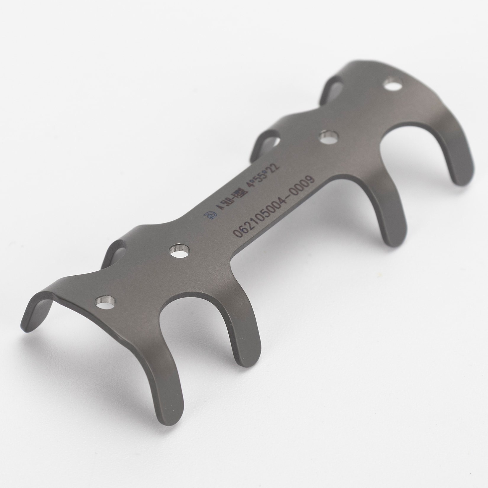
Система коррекции воронкообразной деформации грудной клетки
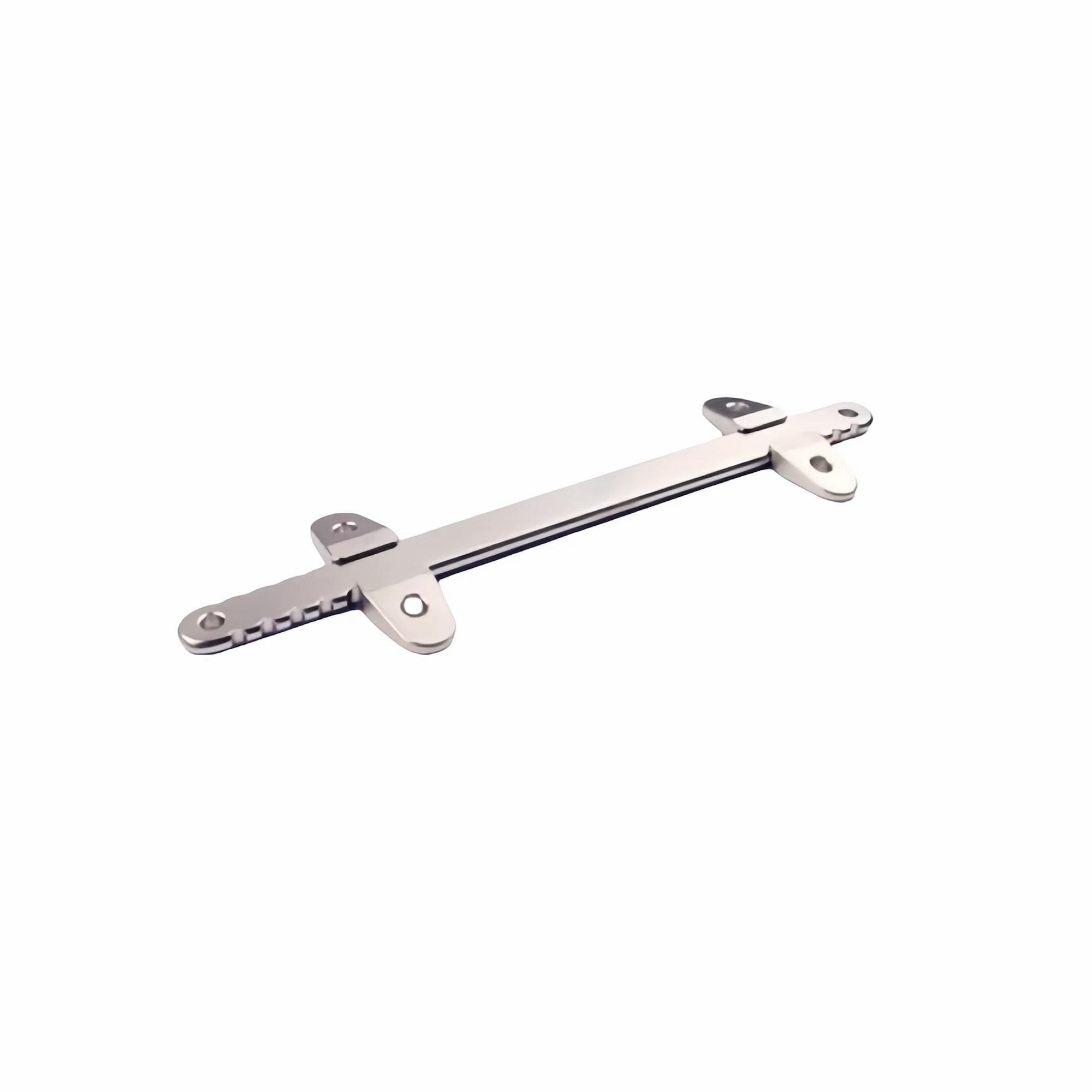
Система коррекции воронкообразной деформации грудной клетки подходит для коррекции и лечения врожденной воронкообразной деформации грудной клетки у детей или при рецидиве воронкообразной деформации грудной клетки у детей.
Артикул № :
GOHE059минимальный заказ :
10 PiecesКлассификация :
Class IIIЦвет :
No Color/Custom ColorИсточник :
Xiamen, ChinaОплата :
T/T 50% and balance before shipmentВремя выполнения :
Depends on the order circumstancesСистема коррекции воронкообразной деформации грудной клетки
The Хирургическая система Pectus Excavatum Состоит из ортопедической пластины (модель JXB01) и фиксирующей пластины (модель GDB01). Ортопедические и фиксирующие пластины доступны в различных спецификациях в зависимости от размера. Изделие изготовлено из чистого титанового материала TA3G, соответствующего стандартам GB/T 13810. Поверхность изделия неокрашенная, упаковка нестерильная.
Технические характеристики системы хирургической коррекции Pectus Excavatum:
Таблица 1: Технические характеристики ортопедической пластины (единица измерения: мм)
| Описание | Модель | Длина | Ширина | Толщина | |
| Ортопедическая пластина | JXB01 | 178, 190, 203, 216, 229, 241, 254, 267 | 12 | 3 | |
| 279, 292, 305, 318, 330, 343, 356, 368, 381, 394, 406 | 13 |
Таблица 2: Технические характеристики фиксирующей пластины (единица измерения: мм)
| Описание | Модель | Длина | Ширина | Толщина | Глубина паза | Ширина слота |
| Фиксирующая пластина | GDB01 | 50 | 15 | 5 | 3 | 12 |
| 60 | 16 | 12 | ||||
| 60 | 16 | 13 |
Инструкции к устройству для коррекции воронкообразной деформации грудной клетки:
1. Операция проводится под общим наркозом с интубацией трахеи;
2. Мягкой сантиметровой лентой измеряется длина двусторонней средней подмышечной линии на грудной стенке. Выбирается ортезная пластина такой же длины или на 1-2 см короче. Ортезная пластина формируется с помощью формовочных щипцов в соответствии с формой грудной стенки пациента;
3. Продольный разрез выполняется между двусторонней передней подмышечной линией и средней подмышечной линией в самой нижней точке грудины. Торакоскоп вводится в правую сторону разреза, на 1-2 межреберья ниже разреза. Под торакоскопическим контролем разделитель тракции проводится через самую высокую точку межреберья в правую грудную полость, а затем проводится по самой низкой точке грудины в левую грудную полость, выходя через левый разрез. Тяговый ремень фиксируется к разделителю тракции. Следуя первоначальному пути разделителя тракции, ремень тракции проводится из левого разреза в правую сторону, разделяя ремень тракции и разделитель тракции. Ремень тракции фиксируется к ортезной пластине, и под руководством ремня тракции вогнутая сторона ортезной пластины проводится из правого разреза в левую сторону. Ортезная пластина поворачивается на 180° с помощью вращающейся ручки;
4. Оба конца ортопедической пластины вставляются в пазы фиксирующей пластины. Фиксирующая пластина помещается на ребра, и фиксирующая пластина пришивается к надкостнице ребер с помощью нерассасывающихся швов. В качестве альтернативы может быть применен метод фиксации, который хирург сочтет подходящим на основе соответствующей литературы;
5. Разрезы с обеих сторон пациента зашиваются хирургическими нитями.
Система коррекции воронкообразной деформации грудной клетки подходит для коррекции и лечения врожденной воронкообразной деформации грудной клетки у детей или при рецидиве воронкообразной деформации грудной клетки у детей.
Подробности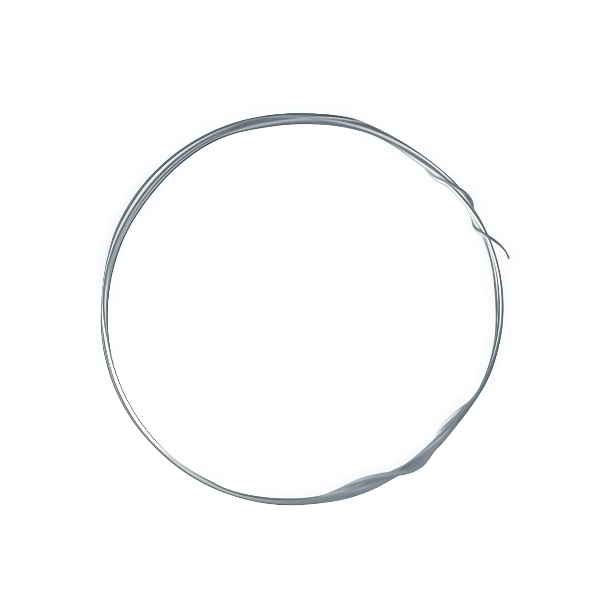
Гибкая проволока из нержавеющей стали применяется для внутренней фиксации переломов конечностей.
Подробности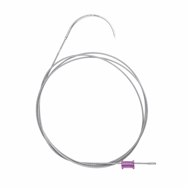
Медицинское устройство для фиксации грудины после торакотомии, способствующее заживлению и стабильности.
Подробности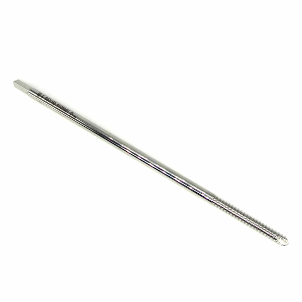
Применяется для вытяжения или внутренней фиксации переломов конечностей во время репозиции переломов.
Подробности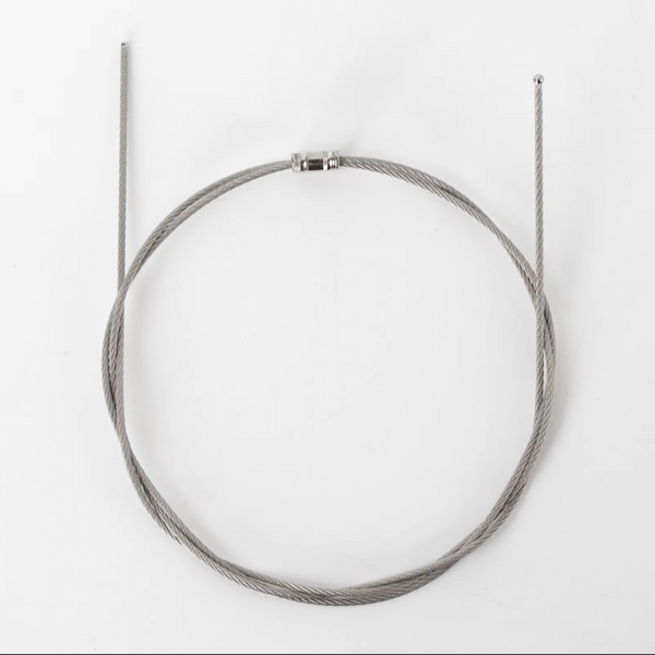
Подходит для интрамедуллярной фиксации и связывания переломов конечностей.
Подробности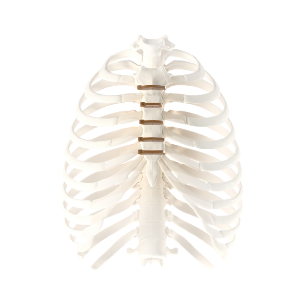
Лигатура для грудины из ПЭЭК обеспечивает быстрый метод закрытия грудной клетки, обеспечивая при этом постоянную и стабильную фиксацию грудины.
Подробности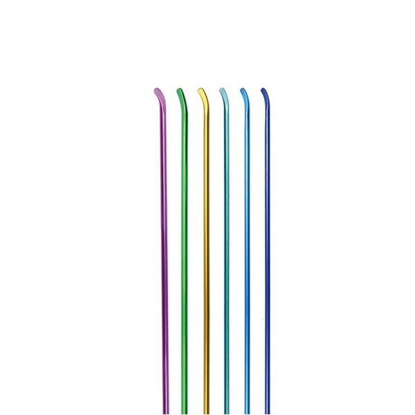
Эластичный интрамедуллярный стержень является безопасным и эффективным средством фиксации переломов, особенно подходящим для лечения переломов длинных трубчатых костей у детей и подростков.
Подробности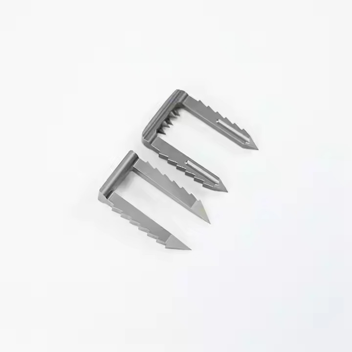
Устройство, используемое в ортопедической хирургии для фиксации переломов и восстановления костей.
Подробности
Подпишитесь на нашу рассылку, чтобы получать обновленную информацию, рекламные акции и идеи.


 Поддерживается сеть IPv6


 Получить цену